Fetal pig dissection labeled pictures serve as an invaluable educational tool, providing a detailed visual guide to the anatomy and physiology of this essential species. This article delves into the fascinating world of fetal pig dissection, examining its historical significance, ethical considerations, and practical applications.
The meticulous labeling of structures in these images empowers students and researchers to identify and understand the intricate components of the fetal pig’s anatomy, facilitating a deeper comprehension of its biological systems.
Fetal Pig Dissection Overview
Fetal pig dissection is an integral part of science education, providing students with a unique opportunity to explore the anatomy and physiology of a developing mammal.
It enhances students’ understanding of the principles of biology, including organ systems, embryonic development, and comparative anatomy. By dissecting a fetal pig, students gain hands-on experience in scientific inquiry, critical thinking, and problem-solving.
Ethical Considerations
Fetal pig dissection raises ethical concerns regarding the use of animals in education. It is crucial to ensure that animals are treated humanely and that their use is justified by the educational value of the dissection.
Ethical guidelines for animal dissection have been established by organizations such as the National Science Teachers Association (NSTA) and the American Veterinary Medical Association (AVMA). These guidelines emphasize the importance of obtaining animals from reputable sources, minimizing their suffering, and disposing of them responsibly.
History of Fetal Pig Dissection
Fetal pig dissection has been used in science education for over a century. It was first introduced in the late 19th century as a way to teach human anatomy and physiology. Over time, it became a standard practice in high school and college biology courses.
Today, fetal pig dissection remains a valuable educational tool, despite the development of alternative methods such as virtual dissection and computer simulations.
External Anatomy
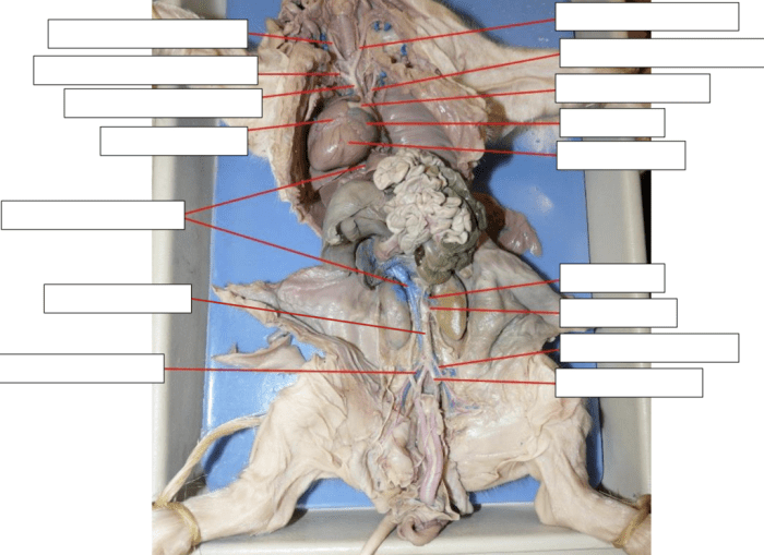
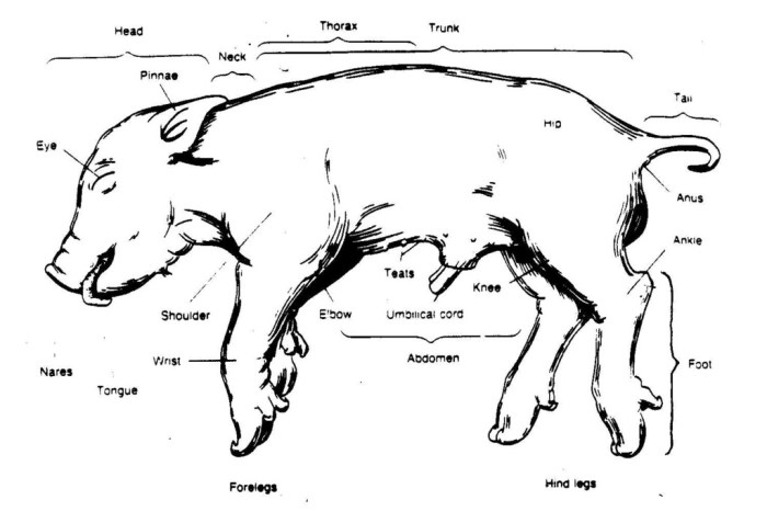
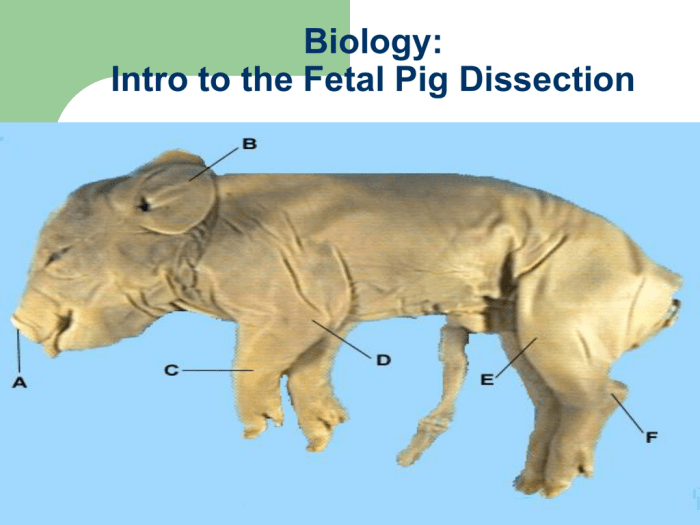
The external anatomy of a fetal pig provides insights into its overall morphology and adaptations. By examining various structures, we can understand the pig’s body plan, locomotion, and sensory capabilities.
The following table Artikels the key external structures of a fetal pig, along with their locations, functions, and relevant anatomical terms:
| Structure | Location | Function | Anatomical Terms |
|---|---|---|---|
| Head | Anterior end | Contains sensory organs, brain, and mouth | Snout, eyes, ears, nostrils |
| Neck | Connects head to body | Provides flexibility and supports the head | Cervical vertebrae |
| Trunk | Central region | Houses internal organs and provides support | Thoracic cavity, abdominal cavity |
| Limbs | Anterior and posterior ends | Enable locomotion and support | Forelimbs (arms), hindlimbs (legs) |
| Tail | Posterior end | Assists in balance and communication | Caudal vertebrae |
| Umbilical cord | Anterior abdomen | Connects the fetus to the placenta | Amniotic sac, placenta |
Illustrations/Images of External Anatomy
Detailed illustrations or images of the external anatomy of a fetal pig can provide a visual representation of the structures discussed above. These images can enhance understanding and aid in the identification of specific features.
Internal Anatomy
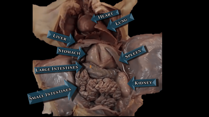
The internal anatomy of a fetal pig is complex and comprises various organs and systems that perform essential functions. Understanding the internal structures provides insights into the development and physiology of the animal.
The major internal organs of a fetal pig can be classified into the following categories:
Respiratory System
- Lungs:Spongy, air-filled organs located in the thoracic cavity. They facilitate gas exchange between the blood and the environment.
- Trachea:A tube-like structure that connects the lungs to the pharynx. It allows air to pass into and out of the lungs.
- Bronchi:Primary branches of the trachea that lead to the lungs. They divide into smaller bronchioles within the lungs.
Cardiovascular System, Fetal pig dissection labeled pictures
- Heart:A muscular organ located in the thoracic cavity. It pumps blood throughout the body, providing oxygen and nutrients to tissues.
- Arteries:Blood vessels that carry oxygenated blood away from the heart to various organs and tissues.
- Veins:Blood vessels that carry deoxygenated blood back to the heart.
Digestive System
- Esophagus:A muscular tube that connects the pharynx to the stomach. It transports food and liquids swallowed.
- Stomach:A J-shaped organ that secretes digestive enzymes and acids to break down food.
- Small Intestine:A long, coiled tube where most of the digestion and absorption of nutrients occur.
- Large Intestine:A shorter, wider tube that absorbs water and electrolytes from undigested food.
Urinary System
- Kidneys:Bean-shaped organs that filter waste products from the blood and produce urine.
- Ureters:Tubes that carry urine from the kidneys to the urinary bladder.
- Urinary Bladder:A sac that stores urine before it is expelled through the urethra.
Dissection Procedures
Fetal pig dissection involves a systematic approach to examine the internal and external anatomy of a developing mammal. To ensure a successful and safe dissection, follow these step-by-step guidelines and adhere to the specified safety precautions.
Step 1: Preparing the Workspace
1. Gather necessary materials: dissection tray, fetal pig specimen, dissecting tools (scalpel, scissors, forceps, probes), gloves, and safety goggles.
2. Wear appropriate personal protective equipment (PPE), including gloves and safety goggles, throughout the dissection.
3. Set up the dissection tray in a well-lit and ventilated area.
Step 2: External Examination
1. Examine the external features of the fetal pig, including the head, neck, limbs, tail, and body cavity.
2. Identify and locate external structures such as the eyes, ears, nose, mouth, umbilical cord, and genitals.
3. Make superficial incisions to expose underlying structures, such as the abdominal and thoracic cavities.
Step 3: Internal Examination
1. Using a scalpel, carefully cut through the abdominal wall to expose the abdominal cavity.
2. Identify and locate internal organs, such as the stomach, liver, intestines, kidneys, and bladder.
3. Remove organs for closer examination and to study their structures and relationships.
Step 4: Thoracic Examination
1. Cut through the thoracic wall to expose the thoracic cavity.
2. Identify and locate the heart, lungs, and trachea.
3. Study the structures of the heart and lungs, including their chambers and blood vessels.
Step 5: Cleaning and Disposal
1. After the dissection is complete, thoroughly clean all tools and equipment used.
2. Dispose of the fetal pig specimen and any biological waste according to laboratory protocols.
Troubleshooting Common Dissection Challenges
1. Difficulty finding structures:Use a probe or forceps to gently separate and identify structures.
2. Damage to organs:Use sharp instruments and handle organs with care.
3. Excessive bleeding:Use forceps to clamp blood vessels and apply pressure to stop bleeding.
Educational Applications: Fetal Pig Dissection Labeled Pictures
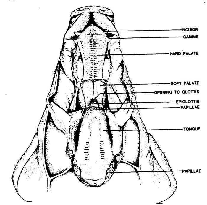
Fetal pig dissection serves as an invaluable educational tool, fostering a profound understanding of anatomy, physiology, and embryology. Through hands-on exploration, students gain an intimate appreciation for the intricate workings of living organisms.
Dissection allows students to visualize and comprehend the spatial relationships between organs, muscles, and other structures within the body. It enhances their ability to identify and differentiate various anatomical features, contributing to a comprehensive understanding of the human body.
Embryology
Fetal pig dissection provides a unique opportunity to study embryological development firsthand. Students can observe the various stages of organ formation, gaining insights into the complex processes that shape life before birth.
Alternative Dissection Methods
Fetal pig dissection is a traditional method of anatomy education, but alternative methods like virtual dissection and computer simulations are gaining popularity. These methods offer advantages and disadvantages that educators should consider.
Advantages of Virtual Dissection and Computer Simulations
- Accessibility:Virtual dissection software and computer simulations are accessible from anywhere with an internet connection, making them ideal for remote learning or students with mobility limitations.
- Convenience:Virtual dissections can be paused, rewound, and repeated as needed, allowing students to learn at their own pace and focus on specific anatomical structures.
- Ethical implications:Virtual dissection eliminates the need for animal dissection, raising ethical concerns about the use of animals in education.
Disadvantages of Virtual Dissection and Computer Simulations
- Lack of hands-on experience:Virtual dissections and computer simulations do not provide the same hands-on experience as traditional dissection, which is crucial for developing spatial reasoning and dexterity.
- Cost:Virtual dissection software and computer simulations can be expensive, especially for schools with limited budgets.
- Technical limitations:Virtual dissections and computer simulations may have technical limitations, such as latency issues or limited resolution, which can affect the learning experience.
Ethical Implications of Using Alternative Dissection Methods
The use of alternative dissection methods raises ethical considerations. Some argue that virtual dissection and computer simulations can reduce the number of animals used in education, while others believe that hands-on dissection is essential for a comprehensive understanding of anatomy.
Educators must carefully weigh these ethical implications when choosing the most appropriate dissection method for their students.
Quick FAQs
What are the ethical considerations involved in fetal pig dissection?
Fetal pig dissection involves the use of animal specimens, which raises ethical concerns. It is crucial to ensure that the pigs are obtained from reputable sources, have been treated humanely, and are euthanized in a humane manner.
What are the educational benefits of fetal pig dissection?
Fetal pig dissection provides hands-on experience in anatomy and physiology, allowing students to visualize and manipulate the structures they are studying. It enhances their understanding of the complex relationships between different organs and systems.
What are some alternative dissection methods to fetal pig dissection?
Alternative dissection methods include virtual dissection software, computer simulations, and 3D models. These methods offer advantages such as the ability to zoom in and out, rotate structures, and explore anatomical relationships in an interactive manner.